Consequences of knee injuries

The increased frequency of knee injuries is due, on the one hand, to the complexity of its anatomical structure, and on the other hand, to the constant increased load during walking and standing. The knee joint is the main supporting structure of the body. Trauma means damage to the soft tissues and bone structures of the joint itself. Dangerous in terms of the consequences of falling from a height. Then the internal integrity of the knee joint with all its ligaments and kneecap is damaged. They will remind of themselves with swelling, pain – both acute and aching. The main consequence of a knee injury is limited movement due to joint stiffness. Kinds There are 3 main groups of knee joint injuries. – Damage to the bone structures of the joint in the form of fractures. – Soft tissue injuries, consequences of sprains, ruptures and tears of ligaments, meniscus. – Combined injuries. Systematization of damage: – A bruise is the mildest type of injury. – Sprains of the tendons and ligaments that support the kneecap. – Meniscus rupture – occurs when the knee is turned sharply with a fixed foot. This type of injury always requires surgical intervention. – Complete and incomplete ruptures of the knee ligaments – the result of forceful impacts on the knee: traffic accidents, sports, falls, etc. – Cartilage damage – occurs with bruises or intra-articular fractures. – Fractures or cracks in the knee bones. – Dislocation of the kneecap – an injury usually seen in adolescents, at the age of 13-18 years. Symptoms and signs General symptoms of injuries: – swelling; – limited mobility, inability to perform certain movements; – crunching, clicking; – increased temperature in the area of injury; – inability to lift a heavy object; – acute pain – occurs a few seconds after the injury; – constant aching pain; – decreased sensitivity due to damage to nerve endings; – hemarthrosis – accumulation of blood in the joint cavity. Treatment methods If we are talking about a minor injury, treatment is usually limited to conservative. It includes therapeutic exercises, physiotherapy and medication. The choice is determined by the doctor. Knee injuries are usually painful, so analgesics based on Ibuprofen, Ketoprofen, etc. are prescribed without fail. Treatment of ligament and meniscus injuries always requires immobilization of the joint by applying a plaster splint or orthosis to the entire leg. In case of bruises, the splint or plaster is removed after 1-2 weeks, in case of ligament ruptures – after 2 months. In case of ligament ruptures, surgical treatment is prescribed in the form of ligament suturing or plastic surgery. Physiotherapeutic measures will be mandatory – magnetic therapy, ultrasound, shock wave therapy, electrophoresis, dynamic currents, UHF, electromyostimulation and lymphatic drainage massage of the thigh, paraffin applications. Today, knee injuries are treated with arthroscopy instead of traditional surgery. This is the standard of leading clinics around the world. Only well-equipped Hospitals like Evercare and Specialist like Dr Waqas Javed can handle this type of treatment. Instead of a traditional incision, the doctor makes two tiny holes that are used to insert surgical instruments and an arthroscope. As a result, the recovery period is significantly reduced. Such patients can be discharged within 24 hours because they immediately begin to walk without crutches and plaster. Results The prognosis is always better with early treatment. Even severe injuries can generally be cured in 2-3 months of intensive therapy. Partially damaged ligaments can fully recover in 1-1.5 months. A complete ligament rupture will require a rehabilitation period of up to a year. Rehabilitation and lifestyle restoration Rehabilitation is no less important in knee treatment. The surgeon, simply put, will “assemble” the joint, restore its anatomy or replace it with an artificial analogue, and then rehabilitation is needed. It provides 50% success after any operation, no matter how high-tech it is. This includes doing exercises under the guidance of a specialist, kinesiotaping, training on special exercise machines. The rehabilitation itself includes 3 stages: – Restoration of the range of motion of the knee, at first the knee joint is moved “passively”, forcibly. – Restoration of tone, strength of muscle tissue, compensation of fiber atrophy. – Work on restoring the gait stereotype, correct dynamics of movements, distribution of load on damaged tissues. All stages must be carried out under the supervision of specialists – from an orthopedic doctor to rehabilitation specialists. Lifestyle for knee injuries The further way of life implies strengthening of muscles, wearing knee pads. Protective devices in the form of orthoses, bandages fix and stabilize the joint. The victim should continue to use the bandage for a certain period of time, even if he feels healthy, at least until the doctor confirms it himself. The goal of knee treatment and recovery is to return to the previous level of activity and quality of life.
Consequences of elbow injuries

The consequences of elbow injuries can be quite serious. The most common complication is contracture – a disorder of motor activity. Joint mobility is impaired as a result of scar and fibrous formations in the tendon area. Another consequence may be joint instability. This problem occurs with ligament damage, dislocation of the radial head and forearm. A similar complication develops if the distal area of the biceps tendon becomes inflamed. Experienced doctors working in the clinic quickly diagnose complications. The medical center has its own diagnostic base. All examinations – CT of the elbow joint , MRI of the elbow , ultrasound can be done within the walls of the clinic, get qualified medical advice, start treatment in a timely manner, which will definitely give results. Kinds The most common elbow injuries include: – bruises; – sprains; – fractures; – dislocations. Bruises are the result of blows and falls. Sprains occur when the integrity of the ligament fibers is partially or completely disrupted. Dislocations are classified as isolated and pronation. Fractures can be open or closed, with or without displacement. Symptoms and signs The main signs of an elbow injury are: – Sharp pain that occurs at the moment of injury. – Numbness and tingling in the area of the hand and forearm. – Swelling in the area of the affected joint. If left untreated, discomfort in the elbow area can become chronic. When a large amount of blood accumulates, a hematoma appears. The pain may increase when moving the limb, raising and lowering the arm. Some people find it painful to move their wrist and fingers. They lose the ability to fully bend and straighten the elbow. Numbness and tingling are caused by damage to blood vessels and nerves. In some cases, the mobility of the injured arm may be abnormal. There are uncharacteristic movements in the elbow, the arm bends unnaturally. Treatment methods Therapy for elbow joint injuries is performed simultaneously in several directions. To relieve pain and swelling, the patient is prescribed nonsteroidal anti-inflammatory drugs and painkillers. Local compresses help well in such cases. If the tablets are ineffective, injections are prescribed. Simultaneously with drug treatment, immobilization of the injured arm is performed – the limb is fixed with an orthosis or a plaster cast is applied. After the bones have grown together and the injured joint has been restored, rehabilitation is carried out, including physiotherapy and therapeutic massages, physical education with special exercises that improve mobility and restore the range of motion. Results The results of treatment largely depend on early diagnosis, the nature of the injury and the chosen method of therapy. The earlier the course is started, the greater the chances of avoiding negative consequences. The patient must follow all medical instructions, in which case one can expect full restoration of elbow mobility after the injury. Rehabilitation and lifestyle restoration During the rehabilitation period after elbow injuries, it is necessary to perform physiotherapy procedures, do a warm-up before intense physical activity. As needed, use special ointments, fixing bandages and dressings during training. Minor bruises heal in a couple of weeks. Recovery from a complex injury takes much longer – from six months or more. The rehabilitation period largely depends on the patient’s age. At a young age, recovery processes are faster. Elderly people need more time. Lifestyle for Elbow Injuries If you have an injured elbow, you need to be careful when moving, especially if the street is icy and there is a high risk of falling. The diet should include foods rich in calcium and phosphorus, and drink vitamin complexes enriched with microelements that increase bone strength and improve the condition of joints. The consequences of an elbow injury rarely end in disability, but people who play sports professionally have to end their careers. The rest are forced to suffer from limited mobility of the elbow joint, which significantly reduces the quality of life.
Hemorrhage in the knee joint

Hemorrhage into the knee joint is called hemarthrosis. It manifests itself as pain, swelling, decreased mobility and changes in the contours of the joint. Blood from the knee must be evacuated, and this requires a puncture of the joint. Subsequently, the underlying disease that caused the hemorrhage is treated. Causes of hemorrhage into the joint cavity Depending on the cause of occurrence, knee hemorrhages are divided into three groups: Traumatic. Non-traumatic. Postoperative. The most common type of bleeding into the joint cavity is traumatic hemarthrosis. For blood to leak into the joint in significant quantities, serious tissue damage must occur: cartilage, bones or ligaments. Therefore, bleeding into the joint is a sign of severe trauma that may require surgical intervention: Up to 70% of all knee bleeds in adults are due to anterior cruciate ligament (ACL) rupture. In 10% of cases, the cause of hemorrhage is a meniscus tear. In 2-5%, hemorrhage is caused by a torn cartilage fragment or an intra-articular bone fracture. In 5% of cases, the cause is a rupture of the posterior cruciate ligament or knee joint capsule. In children, the leading cause is lateral patellar dislocation. Non-traumatic hemorrhage is usually associated with a blood clotting disorder, such as hemophilia, vitamin K deficiency, liver cirrhosis, and anticoagulant use. Rare causes of non-traumatic hemorrhage in the knee joint include diabetic arthropathy, vitamin C deficiency, septic arthritis, tumors in the knee area, hemangiomas, as well as ruptures of the arteries of the knee joint due to osteoarthrosis and degenerative changes in the posterior horn of the medial meniscus. Postoperative hemorrhages are most often associated with endoprosthetics. Less often, it develops as a complication of other knee surgeries. Symptoms After an injury, the joint swells over several hours. Mobility is significantly reduced, but the pain is not always severe. The pain can range from mild to severe, depending on the nature of the injury, and usually increases as blood accumulates inside the joint due to the pressure it exerts. All other symptoms depend on the type of injury. For example, when the meniscus is torn, the knee joint is blocked: a person cannot straighten, or less often bend, the leg. When the anterior cruciate ligament is torn, a feeling of instability appears in the knee, and during a clinical examination, the doctor can determine a positive anterior drawer symptom: the shin is excessively displaced forward during passive movement, since the tibia is no longer held by the torn ligament. Diagnostics Using ultrasound, you can see fluid in the knee, but you cannot reliably determine that it is blood and not inflammatory exudate. The most accurate method is considered to be MRI. With this procedure, it is possible to distinguish blood from other fluids, as well as to establish the cause of the hemorrhage, for example, to visualize torn ligaments or menisci. When intra-articular bone fractures are suspected, which account for only a small proportion of knee hemorrhage cases, radiography and CT scanning are the preferred diagnostic options. Treatment of bleeding in the knee joint Patients with hemarthrosis require a puncture of the knee joint. The doctor performs this procedure under local anesthesia. The doctor inserts a needle into the joint cavity to evacuate the accumulated fluid and inject medications into the knee. This procedure quickly reduces pain and improves knee mobility by reducing intra-articular pressure. The color of the fluid that the doctor obtains from the knee can help the doctor guess the cause of the hemorrhage. It can be pink, red, or brown, and in severe trauma it may contain fatty droplets. Other methods of conservative treatment: ice; anti-inflammatory drugs; immobilization; compression bandages. During the recovery period, massage, exercise therapy, and physical therapy are used. Some patients will need surgery to partially remove the meniscus or reconstruct the anterior cruciate ligament. At Dr. Waqas Javed’s Clinic, all knee interventions are performed using a minimally invasive arthroscopic method. Possible complications Severe or recurrent hemarthrosis can lead to destruction of intra-articular cartilage and osteoarthritis. The toxic effect of blood on the articular membrane causes its hypertrophy (proliferation) and fibrosis (scarring). Most often, complications develop with bleeding into a joint against the background of hemophilia, but this is a rare cause, accounting for no more than 1% of cases, and is mainly diagnosed at the age of 2-3 years. Repeated episodes of bleeding into the joint cavity can cause arthropathy with impaired joint mobility and patient disability. This complication develops in 20% of patients with hemophilia. In addition to the knee, other joints are also affected. Rehabilitation and exercise therapy After surgery or a period of immobilization, patients require rehabilitation. How long it will take depends on the nature of the injury. If after meniscus resection recovery takes only one and a half months, then after a rupture of the anterior cruciate ligament and its reconstruction, recovery takes six months. The main means of rehabilitation is considered to be therapeutic exercise, but other procedures are additionally used, such as massage, physiotherapy, mechanotherapy, electromyostimulation. Forecast After a knee puncture, the pain goes away in a few days, and most other symptoms disappear completely in a few weeks, unless there are gross anatomical defects in the knee. If there are such defects, then without treatment, a person will suffer from joint instability or recurrent blockades. Inflammatory processes (reactive synovitis) will worsen, and in the long term, the risk of osteoarthritis increases. Surgical treatment may be required to prevent these consequences. Prevention Prevention of knee hemorrhages is identical to injury prevention. It is necessary to warm up before training, exercise in good shoes, and avoid running on slippery surfaces. Unfortunately, there are no measures that will protect against knee injury with a 100% guarantee. If you notice that your knee is swollen, painful, and difficult to move as a result of an injury, contact Dr. Waqas Javed in Lahore. We will conduct diagnostics and find out whether there is blood in the joint or not. If there is, we will perform
Exercises after knee meniscus surgery
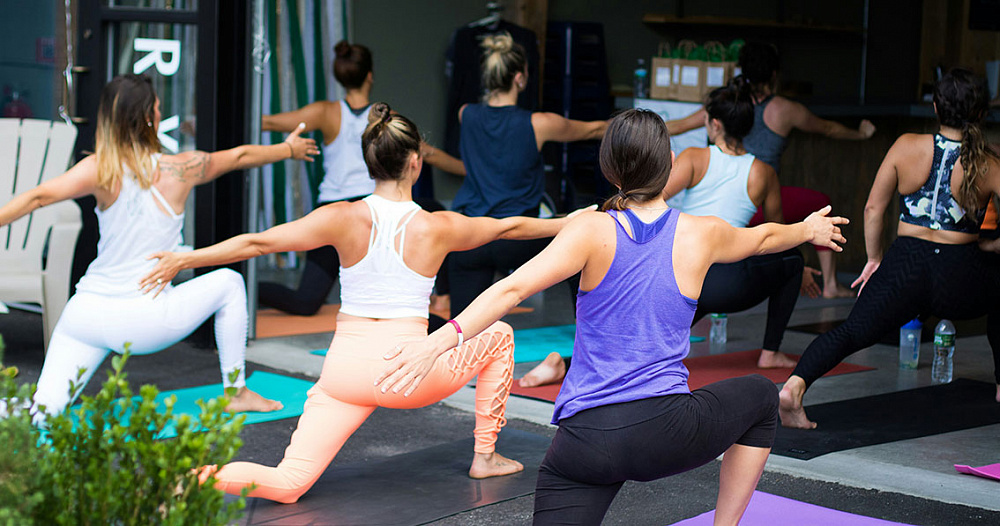
Physical therapy is the most important part of rehabilitation after any operations on the structures of the musculoskeletal system. Although damaged tissues heal on their own, it is important to do exercises to restore muscle strength and range of motion in the joint. The complex is selected individually for each patient. At Dr. Waqas Javed’s Clinic, you can work out with a personal physical therapy instructor. General recommendations before starting training At Dr. Waqas Javed’s Clinic, all surgeries for knee meniscus injuries are performed using arthroscopy. Arthroscopy is a minimally invasive technique that involves performing the intervention through several punctures. Therefore, after you have had a partial meniscus removal, recovery will not take long. It will be faster than after other surgeries. In a week or two you will be able to walk without crutches, and in a month and a half you will feel healthy. In three months you will return to the previous functional state that was before the meniscus injury and surgery. The exercise program begins 2-7 days after surgery. This approach varies from clinic to clinic. The time to start training also depends on the patient’s condition and the specifics of the surgery performed. Before beginning any exercise involving the knee, it is important that most of the swelling has gone down and that the patient is not in severe pain, although some mild discomfort in the operated limb is possible. When choosing exercises, determining the intensity and frequency of training, the general condition of the patient is assessed, the rehabilitation phase is taken into account, as well as the actual condition of the knee joint. What to pay attention to during exercise You should pay attention to your own feelings. If any exercises cause pain or discomfort, tell the instructor about it. At the same time, you shouldn’t be afraid to do exercises even in the first days after surgery. Your knee is not as fragile as you might think. Even if you do something wrong, it doesn’t mean that the tissues inside the knee won’t heal or that you’ll have to have another surgery. Therefore, you should train with sufficient intensity, but at the same time avoid exercises that are not yet acceptable given the rehabilitation phase. For example, it is necessary to choose the right time to add dynamic exercises to isometric exercises, introduce exercises with an expander, with weights, with axial load on the knee joint, etc. An experienced instructor knows which exercises will be useful in a particular period of rehabilitation. Just follow his advice, and the recovery will go smoothly, with good functional results. Exercises in the first 3-7 days after surgery During the first week, you can only walk with support devices such as crutches. Exercises that involve bending the knee joint are not allowed for several days. In the first 3-4 days after the operation, the patient is shown isometric exercises. This means that he tenses the muscles to maintain their tone and improve blood circulation. Examples of such exercises that do not involve the knee joint: A man sits on a bed. He lifts his leg off the floor and moves it 30 cm to the side. Then he returns it to the starting position. Lying on the bed, the person raises the straight leg 15 cm, holds it for 10 seconds, and then lowers it. Since immobilization of the knee joint is not required during the rehabilitation period after meniscus resection, dynamic exercises can be done after 4-5 days, if they do not cause severe discomfort. Examples of such exercises: A person lies on a bed. He bends his leg and pulls his heel towards himself. Over time, this exercise can be made more difficult: not only pull the heel towards yourself, but also lift it. The man sits on the edge of the bed. He bends his knee and smoothly relaxes the muscles of the thigh. Exercises for 2-4 weeks after surgery From 2-3 weeks, the training possibilities are significantly expanded, as the surgical wounds heal. A person can do the following exercises: Lying on his back, he lifts his leg and rotates it four times. He lowers it and rotates the other leg. He does 10 repetitions on each side. Lying on the stomach, the person bends the legs at the knees and lifts the hip. Lying on his stomach, the patient moves his straight leg back. Lying on your stomach, bring your bent knee to your chest. These are just examples of exercises that are not a complete recovery program. The program can include dozens of types of exercises, and the complex should be selected individually. Exercises for 6-12 weeks after surgery From the sixth week, the functional recovery period begins. You can do almost any exercise, since all the tissues in the knee have already recovered. Now the task is to build up muscles and fully restore joint mobility, if the full range of motion has not yet been achieved. Possible exercises: Lying on his back, a person imitates riding a bicycle. The patient sits on a chair. He makes movements with his legs as if he were swimming breaststroke. A person grasps small objects with his toes and holds them for 5 seconds. You can now do exercises with an expander. It is secured to the lower third of the shin with a loop. The other end is attached to a support. The optimal load (expander resistance level) for most people is medium. Examples of exercises with an expander: A person stands and bends his leg at the knee, touching his heel to his buttock. Bends legs at the knees while lying on stomach. The patient abducts his leg while standing. During the functional period, you can also train with weights, for example, doing shallow squats with a barbell on your back or lifting weights with your legs while sitting on a chair and bending your knees.
Basic methods of diagnosis and treatment of various types of knee pain
Knee pain is a non-specific symptom that develops against the background of a huge number of diseases. You are unlikely to be able to determine the cause of pain while sitting at home on the couch with a smartphone in your hand and reading the Internet. We will tell you about the possible causes of pain and methods for solving the problem, but for diagnosis you will in any case need a medical examination and instrumental diagnostics. To find out the cause of knee pain and get rid of it, you can contact Dr. Waqas Javed’s Clinic. How the knee joint works It is important to know the approximate structure of the knee joint in order to understand what might hurt inside. The knee joint is a movable joint between the thigh and the shin. It is formed by three bones: the tibia, femur and patella. The articular surfaces of the knee are covered with hyaline cartilage. The knee is a synovial joint. It has a capsule, synovial membrane, and joint fluid. The synovial membrane in some people forms large folds that can become pinched and cause pain. There are ligaments inside the knee that keep it stable. Injuries can cause ruptures of the anterior and posterior cruciate ligaments, and the medial and lateral collateral ligaments. Between the articular surfaces of the tibia and femur there are two menisci: medial and lateral. These are cartilaginous pads that act as shock absorbers. There are several synovial sacs in the knee joint area. Some of them can become inflamed and painful. There is also a fat pad in the knee. Sometimes it gets pinched, causing chronic pain. Causes of knee pain There are dozens of known causes of knee pain. They can be acute or chronic, trauma-related or not. There are unilateral pains, when only the left or right knee hurts, as well as bilateral ones. Other joints are sometimes involved in the process. In this review, we will discuss only those types of pain that are non-traumatic in origin. Here are the main reasons why knee joints may hurt: osteoarthritis of the knee joint; patellofemoral arthrosis; chondromalacia patella; arthritis; mediapatellar plica syndrome; Baker’s cyst; Hoffa’s disease (incarceration of the fat pad); intra-articular bodies and Koenig’s disease; quadriceps tendinosis; patellar tendinopathy; bursitis of the knee joint (inflammation of the synovial sacs); Osgood-Schlatter disease. There are also pains that are not associated with damage to the structures of the musculoskeletal system. Neurological pains are caused by diseases of the spine, less often – peripheral nerves. Vascular pains are associated with impaired blood supply to tissues or deterioration of venous and lymphatic outflow, for example, thrombosis of the popliteal vein. Pain can be caused by some systemic diseases that affect several joints. In some people, the knee joints begin to hurt first. Examples: rheumatoid arthritis, gout, psoriasis, systemic lupus erythematosus. Possible consequences of knee pain If your knee hurts, the possible consequences depend on the cause. In the best case, the pain will go away without any health consequences if you provide your knee with long-term functional rest. This happens with some bursitis, tendinopathies, and in adolescents – with osteochondropathy of the tibial tuberosity. In such situations, the only problem is the inability to play sports or do physical work. You have to take a break from training for several months. It is worse when the pain becomes constant and does not decrease even with normal, non-sports activities. The consequence is a decrease in the quality of life. You have to constantly take painkillers, and they have a bad effect on the digestive tract, can cause ulcers and bleeding. Finally, the worst outcome is irreversible damage to the cartilage and bone tissue in the area of the articular surfaces of the tibia and femur. Such damage cannot be restored. You have to have knee replacement surgery. Some diseases that you do not treat for a long time lead to this outcome: for example, an old meniscus tear or the presence of loose bone-cartilage bodies in the joint. After treatment, degenerative processes no longer progress, but the tissues that are severely damaged are no longer restored. Additional symptoms Possible additional symptoms in patients with knee pain: limited knee mobility; morning stiffness; swelling; redness; crunch; feeling of instability; deformation. Diagnostics Diagnosis of knee pain begins with examination and palpation of tissues. Then the doctor can conduct functional tests that help determine or suggest which anatomical structures are damaged. In most cases, instrumental diagnostics will be required to confirm and clarify the diagnosis. This may be X-ray or MRI. Less commonly, ultrasound or CT are used. Treatment of knee pain Treatment may be aimed at reducing pain, as well as eliminating or controlling the disease that caused the pain syndrome. Pain relief methods: taking nonsteroidal anti-inflammatory drugs; knee block (administration of local anesthetics with glucocorticoids); various types of physical therapy, the most effective of which in combating pain are high-intensity laser and shock wave therapy. Possible additional therapeutic methods aimed at reducing pain, controlling the disease, improving joint function: physiotherapy; massage; kinesio taping; intra-articular injections of hyaluronic acid; plasma therapy. Some causes of pain can only be eliminated by surgery: for example, resection of a fat pad that is pinched, removal of a Baker’s cyst, or extraction of a free bone-cartilage fragment from the joint cavity. At Dr. Waqas Javed’s Clinic in Lahore, most surgeries are performed using a minimally invasive arthroscopic method, through incisions several millimeters long. Our clinic specializes in the treatment of joint diseases, so you can count on good long-term results. Prevention You can reduce the risk of knee pain by having regular but not excessive physical activity and maintaining a normal weight. When playing sports, you should wear quality shoes with shock-absorbing soles and exercise on non-slip and soft surfaces.
Joint injury – how to recognize?
Joint injuries include bruises, sprains and ruptures of ligaments, tendons, capsules, menisci, cartilage damage, joint dislocations, intra-articular bone fractures. We tell you which injuries are the most common and what to do if you are injured. Classification of joint injuries Due to the occurrence of joint injuries there are: production; not related to production. In turn, production is divided into industrial, agricultural, construction, transport and others. Non-industrial ones can be household, street, road, non-road transport, sports, school and others. By damage mechanism: direct – the damage is localized where the force was applied (for example, a blow to the knee); indirect – the injury is located far from the area where the force is applied (for example, a twisted leg during an unsuccessful landing after a jump). Depending on the duration of the injury, there are: acute – one-time trauma to the joint; chronic – long-term microtraumatization. By time of patient’s request: fresh; obsolete. The timeframes differ for each type of joint injury. The time of transition to an old injury can range from several days to several months. Sometimes intermediate forms are distinguished: for example, subacute injury. Based on the presence of a wound on the skin, trauma can be: closed – without damaging the skin; open – with a wound on the skin, for example, an open bone fracture. Depending on the number of damaged joints and other tissues, trauma can be isolated, combined (several joints) and combined (damage to joints and other anatomical structures). By severity: mild, moderate, severe injury, and some classifications also distinguish an extremely severe degree. Depending on the presence of complications, joint trauma can be complicated or uncomplicated. Complications are different for each trauma. Symptoms and diagnosis of joint injuries The main universal signs of joint injury: pain; edema; deformation; limitation of mobility. Individual injuries may have specific symptoms, but in general, a diagnosis based on clinical signs is not always possible. Sometimes additional instrumental diagnostic methods are used: CT and X-ray are better suited for diagnosing bone fractures, while MRI is considered the best option for diagnosing soft tissue injuries, including ligaments, tendons, cartilage, menisci, and joint capsule. Knee joint injuries Rupture of the anterior cruciate ligament. Most often, it is torn completely. This is one of the most common injuries in sports. To treat it, you have to do an operation with a subsequent long recovery. And if you do not do the operation, chronic instability of the knee develops and the risk of osteoarthritis increases. Meniscus tear. Along with ACL tear, one of the two most common knee injuries. Menisci are cartilaginous pads inside the knee joint. When they tear, they usually do not heal due to lack of blood supply. In some cases, a torn meniscus can be stitched, but more often the damaged fragment has to be removed. At Dr. Waqas Javed’s Clinic, this surgery is performed using a minimally invasive arthroscopic method, with a recovery period of about one and a half months. Other common knee injuries include: rupture of the lateral ligaments: more often the internal one, less often the external one; rupture of the posterior cruciate ligament (rarely isolated); intra-articular bone fractures; ruptures of cartilaginous fragments; damage to the joint capsule. Ankle Injuries Ligament injuries. The most common type of injury. There are many ligaments in and around the ankle that can be damaged as a result of a twisted ankle. The most frequently injured is the talofibular ligament. A complete rupture leads to a dislocation of the talus. The lateral collateral ligaments are also often torn. Ankle fractures. They account for 60% of all tibia fractures. The main mechanism of injury is the outward rotation of the foot. Subluxation or dislocation of the foot is possible. Often, the deltoid ligament is torn at the same time, as well as the anterior tibiofibular ligament (partial rupture of the tibiofibular syndesmosis). Other possible ankle injuries: fracture and dislocation of the talus; damage to the cartilage of the talus; tendon ruptures. Shoulder joint injuries Shoulder dislocation. The shoulder joint is the most mobile and has almost no ligaments. Therefore, it is the most frequently dislocated joint. Primary dislocation can become habitual due to damage to the anatomical structures that provide stability to the shoulder joint. Bone fractures. They are supratubercular (fractures of the head, anatomical neck), infratubercular (transtubercular, fractures of the surgical neck), and also include fractures and avulsions of the greater tubercle of the humerus. Biceps tendon rupture. Most often occurs against the background of degenerative changes, in men over 40 years old. Usually the tendon ruptures when lifting weights on the biceps (with a bent elbow). Rotator cuff tears also occur, but they are usually degenerative rather than traumatic. Joint capsule damage is possible, but this is an adjunct to shoulder dislocation rather than an independent injury. Elbow joint injuries Bone fractures. Fractures of the bones that form the elbow joint account for 20% of all bone fractures. They can be intra-articular and extra-articular. Intra-articular fractures include fractures of the head, neck of the radius, olecranon and coronoid process. Dislocation. Occurs when falling on a bent arm. The radius and ulna are behind the humerus. Ligament ruptures. The most common are ruptures of the lateral and ulnar ligaments, which become an addition to the elbow dislocation. Causes of joint injury Sports injuries to joints are divided into two groups according to the cause of their occurrence: direct and indirect, which are associated with the characteristics of the human body. Direct causes include poor organization of the training process (weather, footwear, equipment, etc.), excessive loads and lack of medical supervision. Indirect causes include poor physical fitness, poor athletic skills, hidden and established diseases (contraindications to exercise), moral, volitional, disciplinary and other reasons on the part of the athlete. How to recognize a joint injury It is not difficult to recognize a joint injury: if the joint suddenly begins to hurt as a result of a blow, a twisted leg, or the impact of any other one-time factor, and this pain does not
Knee Ligament Injury – Treatment and Prevention
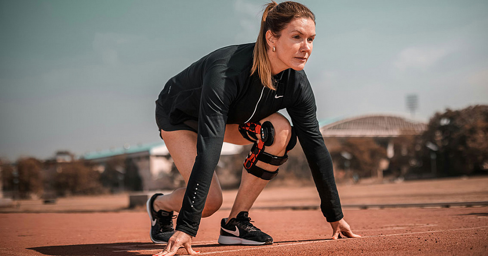
The stability of the knee joint is ensured by its ligaments. They hold the tibia and femur so that the shin does not significantly shift relative to the thigh during physical activity. Knee ligament ruptures are among the most common injuries in sports. If the rupture is complete, then the stability of the joint worsens, and this situation can only be corrected surgically. In case of a partial rupture, conservative treatment tactics can be used. Types of knee ligament injuries The following ligaments can rupture in the knee joint: Anterior and posterior cruciate ligaments (ACL and PCL). Internal and external lateral (collateral). Tears can be complete or partial. While the ACL and PCL are usually completely torn, collateral ligament injuries are more often partial. Therefore, the ACL and PCL have to be reconstructed surgically, while conservative treatment is mainly used for collateral ligament tears. The most common injury is to the ACL. Along with a torn meniscus, this is one of the two most common knee injuries. During an injury, a person can damage both of these structures: the ACL and the meniscus, and it is also possible to combine this with a torn medial collateral ligament. Symptoms Universal symptoms of any knee injury: pain and swelling. These signs are non-specific. They are the same for different injuries, so they cannot serve as a basis for diagnosis. Fluid can accumulate in the knee, which smooths out the contours of the joint. The doctor may discover additional symptoms when examining the patient, palpating tissues and performing functional tests: Collateral ligament damage is characterized by pain upon palpation in the rupture area. An attempt to move the shin to the side opposite to the damage causes sharp pain. Ruptures of the ACL and PCL are manifested by excessive passive displacement of the tibia relative to the femur forward or backward compared to the healthy limb (anterior and posterior “drawer” symptoms). To confirm and clarify the diagnosis, the doctor conducts diagnostics using medical imaging methods. Causes The main causes of ACL and collateral ligament ruptures are sports-related stress. They are often damaged in team sports. Ruptures of the PCL are much less common and are often combined with other knee injuries. More than half of all cases are related to road accidents. Much less often, a rupture of the PCL occurs as a result of a sports or domestic injury. Diagnostics The most effective diagnostic method is MRI. The technique allows determining which ligaments are damaged, whether it is a complete or partial rupture, and which other knee structures are injured. Based on the MRI results, key clinical decisions are made, such as whether a person needs an operation to restore the ligament or whether conservative tactics can be used. Treatment of knee ligament damage Treatment for a partial rupture can only be conservative, while a complete rupture usually requires surgical intervention. If surgery is not performed, the pain will go away over time, but the stability of the knee will deteriorate. There is a feeling of instability when walking, complaints of the knee “flying out”. Over time, osteoarthritis may develop – irreversible destruction of cartilage and bone tissue in the knee joint. To avoid complications, it is better to see an orthopedic doctor in a timely manner and undergo treatment. If the moment for timely treatment is missed, this does not mean that effective treatment is impossible. Reconstruction of a torn ligament can be performed even with an old injury. Conservative treatment Surgery may be avoided in cases of 1-2 degree collateral ligament rupture (partial rupture), but this approach is rarely used in cases of ACL rupture. The main methods of conservative treatment are: immobilization, usually for no more than 2 weeks; cold to the site of injury for the first few days; nonsteroidal anti-inflammatory drugs; massage; physiotherapy; physiotherapy. Suturing the ligament The technique is used only for ruptures of collateral ligaments, if no more than 7 days have passed since the injury. This approach is usually not used for ACL and PCL injuries. A ligament whose structure is preserved can be sutured. If there are signs of fraying, the ligament is reinforced with other anatomical structures, such as the tendon of the semitendinosus muscle. If the lateral ligament is torn from the bone, suture anchors or staple fixation are used to attach it. Ligament reconstruction If more than 10 days have passed since the injury, suturing of the collateral ligament is impossible. In this case, plastic surgery is performed: single-bundle or double-bundle, anatomical or non-anatomical. It can be performed with the patient’s own tissues, donor preserved tissues or synthetic materials such as lavsan tape (lavsanoplasty). Ligament reconstruction essentially means that instead of the old torn ligament, the doctor will create a new one from other tissues. The standard treatment option is autoplasty, for which the anterior, posterior tibial tendon or Achilles tendon is used. In patients who are less demanding of the functional result and are not going to play sports after surgery, lavsan tape can be used for plastic surgery to avoid injury to the donor site. In case of anterior cruciate ligament rupture, the main treatment option is considered to be reconstruction, while ligament suturing is not practiced even in the acute period of injury. Autoplastics are most often used. The material for ligament reconstruction is the patient’s own patellar ligament with a bone block or the tendons of the flexors of the lower leg, which are fixed in bone tunnels. For repeated plastic surgery, the tendon of the quadriceps femoris can be used. Rupture of the PCL is rare, and this injury is usually not isolated, but combined. In case of a complete rupture, plastic surgery with one’s own tissues is required: single-bundle, double-bundle or inlay. The essence of this operation is the same as in the reconstruction of other ligaments: material is taken from the donor site and installed in the knee, fixing it in bone tunnels or fixing the ligament together with the
The harm and benefits of physical activity for the musculoskeletal system

Some say that doing sports is good for you. Others say that it is harmful. Where is the truth? Is it worth doing physical exercises or is it better to sit and lie down more so as not to overexert yourself? Let’s clarify this issue. We will not discuss the benefits of physical exercise for the heart, lungs and other organs. We will focus exclusively on the benefits and harms for the musculoskeletal system. Benefits of Exercise There is no doubt that physical exercise is beneficial for the musculoskeletal system. Therefore, physical education is often used to treat diseases and rehabilitate patients after injuries and operations. This type of training is called therapeutic physical education (LFK). For healthy people, physical training is also useful. Moreover, it benefits all structures of the musculoskeletal system without exception: bones, muscles, joints, cartilage, ligaments, etc. What exactly is the benefit: muscles grow; bones are strengthened; tissue blood circulation improves; ligaments and tendons become more elastic; joint mobility improves (range of motion increases); metabolism in cartilage tissue improves. In addition, a person’s overall physical performance, strength, endurance, speed, coordination, etc. increase. Thus, under the influence of loads, the musculoskeletal system develops, and any development is useful, not harmful. Potential Harm from Exercise They say that physical exercise can wear out joints. In addition, you can get an injury or chronic diseases associated with long-term microtraumatization. In fact, there can be no harm from exercises performed correctly. Only excessive training is harmful – such that it exceeds the body’s current capabilities. These capabilities gradually increase if you experience regular physical activity. Exercise does not “kill” joints. On the contrary: physical inactivity is a risk factor for osteoarthritis – cartilage degeneration. The problem with cartilage tissue is that it is deprived of blood supply. Cartilage can only be nourished from the synovial fluid inside the joint. It is important to constantly “pound” this fluid during physical exercise, since periodic increases in pressure in the joint force nutrients to penetrate into the cartilage, thereby maintaining it in good condition. What can be associated with harm from physical exercise: incorrect technique; too much weight during strength training; too frequent classes; excessively long workouts; lack of warm-up; exercising through pain, through fatigue, through unwillingness to exercise (overtraining). If you know the limits and do the right exercises, you will not be able to get any harm from them. Training brings only benefits. They are recommended to everyone: healthy men and women, patients with chronic joint diseases, children, and the elderly. Even after serious injuries and surgeries, physical training begins literally the next day to speed up recovery. So there are practically no contraindications for doing exercises. The only question is what kind of physical activity you need to get benefits in a particular situation.
Prevention and early diagnosis of degenerative changes in the knee joint in athletes
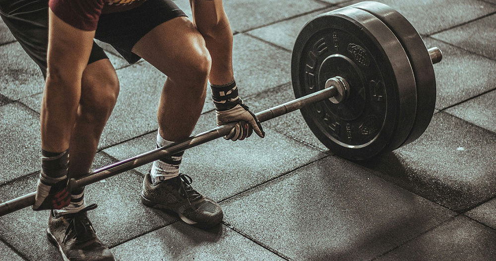
Sports are generally good for your health, but they are bad for your joints. Knees are especially often affected. Athletes get them damaged due to injuries and high-intensity loads. These consequences can be avoided by specifically preventing degenerative changes in the knee joints. What are degenerative changes in the knee joint? Degenerative changes mean that the joints gradually deteriorate. There are tissues inside the knees that are poorly supplied with blood or have no blood supply at all – they are nourished only by synovial fluid. Metabolism in such structures is very slow, so the possibilities for self-recovery are limited. They gradually “wear out” but do not regenerate. As a rule, degenerative changes in the joint are irreversible. Therefore, it is so important to prevent their development. With significant degenerative changes in the knee, it is impossible to restore the meniscus and articular cartilage, and the only radical way to solve the problem is endoprosthetics. Causes of Meniscus Degeneration The risk and rate of degeneration of the menisci and other tissues, such as articular cartilage, depends on several factors: the level of stress on cartilage (in athletes it is usually high); past injuries (most athletes have a history of knee injuries); the condition of cartilage tissue, which depends on congenital characteristics, age, lifestyle (smoking, obesity, endocrine disorders, toxins and other unfavorable factors have a negative effect on the structure of cartilage). The risk of degenerative processes (arthrosis) increases with congenital or acquired deformation of the knee, when the joint is deviated inward or outward. In this case, the load on one “half” of the knee increases. A person’s hyaline cartilage and bones can be destroyed in the places where they contact (the femur and tibia). Also, increased loads in young athletes often lead to degeneration of cartilage tissue in the patellofemoral joint. This is the third component of the knee joint, which is the contact zone of the patella with the femur. Diagnostics To examine patients both for preventive purposes and when complaints from the knees appear, the following methods are used: Clinical examination. The doctor examines the joints, palpates the tissues, evaluates the mobility of the knees, and conducts functional tests. This can identify many diseases and the consequences of injuries, such as a ruptured knee ligament. X-ray. The images can reveal pronounced degenerative changes in the meniscus and articular cartilage of the knee joint, and establish the severity of arthrosis. MRI. The most accurate diagnostic method that helps to detect even the initial, asymptomatic stages of degenerative changes. Not only the destruction of cartilage and bone tissue, bone growths and other obvious problems are noticeable, but also swelling, fraying, changes in the structure of cartilage tissue. Prevention of the development of degenerative changes in the knees of athletes The risk of degenerative changes can be reduced by the following methods: weight loss in case of obesity; quitting smoking, healthy lifestyle; reducing impact loads on the knee joints – running on softer surfaces (rubber, grass, boards, sand instead of asphalt and concrete), as well as using high-quality shoes with shock-absorbing soles; use of orthopedic shoes or insoles; taking dietary supplements, such as chondroitin, glucosamine, collagen; compliance with basic rules that protect against injuries: warm-up, adequate loads, avoiding training when tired, intoxicated, etc. It is important to develop the muscles of the legs, especially the thigh muscles. Strong muscles reduce the load on the articular cartilage. A common cause of degenerative changes in the knees at a young age are untreated injuries. If a person has torn a meniscus, torn a ligament, or suffered another severe injury, and has waited out the acute period, the main symptoms, such as pain and swelling, go away. It seems that there are no consequences for the knee. But in fact, its stability may be impaired, or fragments of cartilage and meniscus may remain in the knee, which constantly injure other anatomical structures. Untreated injuries lead to rapid “wear” of the knee with the need for endoprosthetics after just a few years. Therefore, having received any injury, even if it seems minor, it is worth visiting a doctor to at least undergo diagnostics. It is better to spend 1-2 hours to make sure that everything is ok with the knee than to undergo a complex, traumatic and expensive operation to install an “artificial knee” several years later. Exercises Although exercise causes cartilage degeneration, without exercise, knees also deteriorate because metabolic processes are disrupted. In addition, regular exercise is important for strengthening muscles. Use of orthopedic instruments Orthopedic products help athletes avoid injuries or reduce the load on the knee, thereby reducing the risk of degenerative changes in cartilage tissue. Here are some tools you can use: knee pads; kinesio taping; orthopedic insoles; orthopedic shoes; knee brace. Regular medical examinations of athletes to detect knee problems By playing sports, you fall into the risk group for developing gonarthrosis (degenerative changes in the cartilage of the knee joint). To avoid this disease, special measures are necessary. They are determined individually by an orthopedic doctor. If you play sports and live in Moscow, contact a sports medicine specialist at Dr. Glazkov’s Clinic: once a year, if nothing bothers you; in case of any injury, even if it does not seem serious to you; if you experience pain, a feeling of instability when walking, swelling of the knee, and other symptoms. If there are problems with the knees, it is better to identify and solve them at an early stage. Because late-stage gonarthrosis is difficult to treat, and over time the knee is destroyed so much that an artificial prosthesis has to be installed in its place.
Osteitis, inflammation of bone tissue
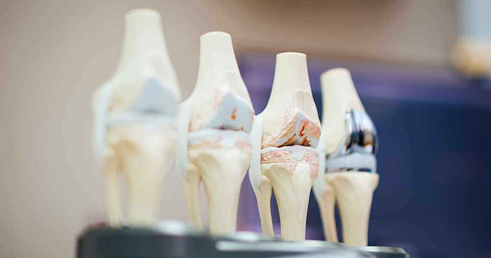
Osteitis is an inflammation of bone tissue. It can be combined with inflammation of soft tissues, periosteum (periostitis) and bone marrow (myelitis). Osteitis can affect any bone in the body. Usually, this inflammation is infectious. The infection can be introduced into the bone from the external environment, during injuries and operations, with the blood flow from other organs, and also directly from nearby inflamed soft tissues. What is this? General information Osteitis is a general medical term meaning that the bone tissue is inflamed. It is sometimes used in dentistry, otolaryngology, but is almost never used in orthopedics. Isolated osteitis is usually tuberculosis of the bones. In all other diseases, inflammation of bone tissue is combined with inflammation of the periosteum and bone marrow. Therefore, in orthopedics and surgery, inflammatory processes of the bone are called osteomyelitis, not osteitis. Causes Osteitis is a disease of microbial origin. Most often it is caused by bacteria, less often by fungi. The causative agents of ostitis can be staphylococci, streptococci, enterobacteria, pseudomonas, E. coli, enterococci, bacteroids, mycobacteria, candida, actinomycetes, Pseudomonas aeruginosa and other flora – both pathogenic and opportunistic. Pathogenesis Osteitis develops as a result of infection getting into the bone. It can get there in different ways, but there are three main ones: trauma with the introduction of infection from the external environment (open fracture, foreign body in the bone, gunshot or shrapnel wound); purulent infection of adjacent soft tissues that gradually reaches the bone; disruption of the blood supply to bone tissue, which most often occurs in diabetes mellitus (diabetic foot); surgery or an invasive procedure, especially one involving the placement of an implant in the bone; infection spreading through the blood from other parts of the body, such as from the lungs in tuberculosis. Osteitis can also develop as a complication after the administration of the BCG vaccine, both as part of vaccination against severe forms of tuberculosis and during treatment. For example, BCG is administered into the bladder to treat cancer of this organ. Classification Depending on the method by which microorganisms enter the bone, ostitis can be: endogenous – bacteria or other microbes that penetrate the bone are inside the body; exogenous – the causative agent of ostitis enters the bone from the external environment, usually as a result of trauma, less often as a result of surgery or a minimally invasive procedure, such as a bone biopsy. Clinical forms: acute osteitis – without bone sequestration according to pathomorphological examination; chronic osteitis – with bone sequesters (islands of bone tissue death) that appear during a long-term inflammatory process. Depending on the localization, ostitis can be of tubular and flat bones. By pathogen: non-specific – caused by non-specific microbial flora; specific – caused by a specific pathogen (tuberculosis, syphilis). Symptoms Symptoms are mild. Many patients have no symptoms at all. If symptoms do appear, the main complaints are mild pain in the bones, which intensifies with movement, hot skin over the bone, swelling, and redness in this area. A slight increase in body temperature is possible. With vertebral ostitis, they become painful when pressed. Complications With ostitis, inflammation can spread: on adjacent tissues – soft tissue structures, joints; to distant organs and tissues – if pus gets into the blood, it can reach the lungs, kidneys, brain and other organs. Osteitis can cause sepsis, thromboembolic complications, and also causes consequences for the musculoskeletal system: bone deformation, fractures. In severe cases, ostitis is treated by amputation of the limb, which leads to patient disability. Diagnostics The following methods are used to diagnose ostitis: MRI. The most accurate non-invasive diagnostic method. Allows to detect signs of ostitis already 3 days after the onset of the disease with a sensitivity of up to 90%. Osteoscintigraphy. An alternative to MRI if there are contraindications. A radioactive substance is introduced into the body, and then during scanning it is checked where it accumulates and in what quantity. The method has high sensitivity, allowing to detect changes in bones in the first days after the onset of the disease. But the specificity is lower than that of MRI. Therefore, osteoscintigraphy is mainly performed on patients who have metal implants in the body (contraindications for MRI). Radiography. The most commonly used method. It is possible to detect foci of demineralization of inflamed bone tissue. The problem is that the changes become noticeable with the loss of more than 20% of bone substance, and become obvious with the loss of more than 50% of bone substance. Therefore, even experienced specialists can detect signs of ostitis on X-rays no earlier than 10 days after the onset of the disease. Blood culture. Helps to identify infectious agents, but only in cases where bacteria enter the bone through the blood – hematogenously. This route of infection is more typical for children, but is rare in adults. Bone biopsy. A mandatory method for confirming the diagnosis. In case of osteitis, doctors must take a tissue sample, examine it under a microscope, and also perform a culture on a nutrient medium to understand which bacteria caused the osteitis and which antibiotics are best to treat the disease. A bone biopsy is performed using an open or percutaneous (minimally invasive) method. An open biopsy is a more accurate method. This is a small operation to remove a fragment of bone tissue. A less accurate, but also less traumatic method is a percutaneous biopsy. This is the insertion of a thick needle, inside which a column of bone tissue remains. At least two samples are taken. The injection must be performed through non-inflamed skin. Otherwise, microorganisms that caused inflammation of the skin, not the bone, may grow on the nutrient medium. Biopsy material culture. An important part of diagnostics, since osteitis can be caused by many pathogens. Based on the culture results, the bacteria that caused the inflammation are identified. Their sensitivity to antibacterial drugs is also assessed. These antibiograms allow choosing the optimal treatment regimen. Treatment Doctors carry out conservative and surgical treatment for