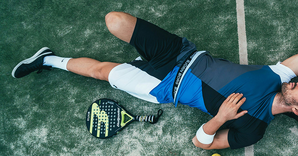
The best sports for joints
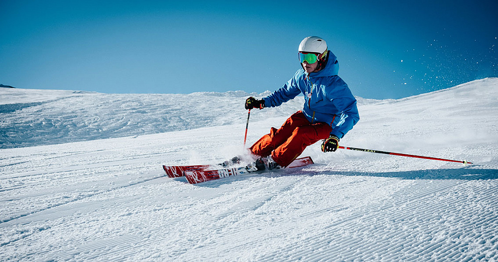
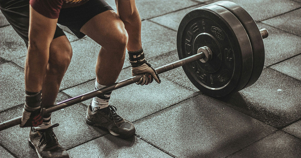
There are over 8,000 types of sports. If you do them for health reasons, and not for outstanding achievements, it is better to prefer those types that are useful or at least not harmful to joints. We tell you what is useful and harmful about sports, how to do it correctly and which types of sports are best for people with problematic joints. Joints and sports Different sports affect joints differently. For example, weightlifting, running, team sports, throwing objects can harm joints. But they cause harm only in the case of very intense, frequent, prolonged loads. If you do sports non-professionally, not often, do not train to exhaustion, do warm-up and cool-down, use high-quality clothes and shoes, then significant harm to joints is unlikely. When doing sports, it is important to increase the load gradually. Do not chase records. Muscles cannot strengthen instantly, and if they are weak, then most of the load falls on connective tissues, including joints. Basic recommendations for playing sports safely for your joints: Adequate loads that increase gradually. When lifting weights, it is better to do more repetitions but with less weight. To reduce the risk of joint problems, you can reduce the range of motion. Example: partial squats. Take breaks from training. This recommendation applies to both strength exercises and cyclic sports. If you feel pain, stop training immediately and take a break for a few days to allow the damaged tissue to recover. When starting to play sports without experience, it is worth using the services of a trainer to gain the necessary skills and knowledge. Listen to your feelings. If you feel that during a certain exercise there is discomfort in the joint, you should change the technique or abandon this exercise. If you are starting to exercise, already having problems with your joints, you should use the services of a physical therapy instructor. Contact Dr. Waqas Javed’s Clinic in Moscow to get help. What kind of sport should people with joint problems do? If there are already problems with the joints, and a person wants to do sports, the first thing to do is to figure out why he needs physical activity. Two main options: To solve the problem with joints. To benefit your overall health and maintain physical fitness without causing any harm to your sore joints. If a person wants to solve a problem with joints, then he needs not sports, but therapeutic physical training. These are special exercises to strengthen the muscles in the area of problem joints. The main goal of therapeutic physical training is not athletic achievements, but the treatment of diseases. It will help reduce pain and restore damaged tissues (for example, tendons). If a person does not use sports as a tool for treating joints, but wants to do it for another purpose, but in such a way as not to harm the joints, then below we offer 7 types of sports that are safe. Walking When a patient is told to limit the load on sore joints, this does not mean that they need to switch to a completely sedentary lifestyle. Lack of physical activity is no less harmful than excessive loads. Walking is a great option to provide moderate loads on joints at a level that will only benefit them. You need to walk on a flat, non-slip surface. It is advisable to use shoes with shock-absorbing soles or individual orthopedic insoles, which can be ordered at our clinic. By creating a moderate load on the joints, walking makes the synovial fluid with nutrients penetrate better into the cartilage tissue, supporting metabolic processes in it. In addition, it strengthens the leg muscles and improves blood circulation in the tissues. Swimming Swimming pool exercises involve all muscle groups, but without axial load on the joints of the lower extremities and spine. Gymnastics Gymnastics is a flexible concept. It can be different. If we are talking about gymnastics for health, then it can be divided into two types: light exercises in the morning (warm-ups) or more intensive and long-term training aimed at increasing muscle strength and endurance: these are static exercises, training on equipment (horizontal bar, parallel bars, etc.), training with objects and weights. Gymnastics, depending on how you do it, can be both harmful to joints and useful. It is also used as one of the methods of treating joint diseases. To get only benefit, avoiding harm, contact Dr. Waqas Javed’s Clinic. Here you can do gymnastics with a personal instructor. He will select a set of exercises for you, depending on which joints hurt, why they hurt, as well as your physical condition and requirements for training results. Elliptical trainer The best cardio option for people with problematic joints. There are no impact loads, like when running, but the muscles and heart work and get stronger. In addition to the usual “vertical” elliptical trainers, there are also “horizontal” ones, on which a person sits during training. Skis Skiing is good for the cardiovascular system, for the muscles of the whole body, and at the same time is at least not harmful to the joints. But we are talking specifically about skiing, and not about a rapid descent from the mountain with the risk of falling and getting injured. Yoga Yoga is a combination of spiritual practices with exercises, mostly static, usually performed without objects and weights. It performs two functions at once: therapeutic physical training to strengthen the muscles in the area of sore joints and a beneficial effect on the psycho-emotional state, which is especially important for patients with a neuropathic component of chronic pain. Pilates Pilates is a type of training that was originally created for people recovering from injuries. These are exercises performed at a slow pace. They provide muscle strengthening, are useful for ligaments and joints, but it is advisable to work with a trainer to get only benefits, without the risk of aggravating the symptoms of the disease. Even people with sore joints can
Joint restoration

Joint restoration is a broad concept that can include a wide variety of processes: rehabilitation after injuries, surgeries, treatment of degenerative diseases, chronic pain, restoration of damaged or destroyed anatomical structures (cartilage, ligaments, tendons) as a result of diseases. Joints can be restored using both conservative and surgical methods. Which ones are required depends on the nature, cause, severity of damage or destruction of the joint, its type, functional state, severity of symptoms and the patient’s requirements for the results of restoration. When joint restoration is required Usually, joints that have been damaged as a result of: injuries; operations; diseases. Recovery after injuries, surgeries and illnesses is called rehabilitation. It is aimed at both anatomical restoration of the tissues that form the joint and functional restoration: increasing muscle strength, range of motion, sensitivity, joint stability and other indicators to the maximum level. If possible, full restoration is ensured: to the level that the person had before the injury or illness. At the same time, the goals of recovery depend not only on the rehabilitation potential. They are agreed upon with the patient and are not necessarily maximum. For example, for older people who do not play sports or do physical work, it is enough to restore the function of the affected limb to the level at which they can normally perform everyday tasks without feeling discomfort or limitations in everyday life. Cartilaginous tissue of joints – features of restoration There is cartilage tissue in the joints. It is destroyed by arthrosis and some other diseases. Many patients want to restore cartilage with medications, physical therapy, exercises, etc. In fact, in most cases, restoration of destroyed cartilage is impossible. This does not mean that treatment is not required. Therapy is necessary at least to prevent the cartilage from being destroyed further. Cartilage restoration is possible only if the cartilage defects are local – formed as a result of trauma or acute disease. Such defects can be closed with fragments of autologous (own) cartilage, which, depending on the size of the defect, is obtained from non-load-bearing sections of the same joint, from another joint or from other tissues. This treatment option is called mosaic chondroplasty. There are also other methods, such as autologous chondrocyte (cartilage cell) transplantation on a collagen membrane or stem cell transplantation. These treatments provide similar results, but are more expensive for the patient. Chondrocyte implantation also requires two surgeries. Arthrosis or arthritis – when joint restoration is needed Arthrosis and arthritis are completely different concepts. Arthrosis is a degenerative process, actually a gradual destruction of the joint. The main reason is the “wear” of the cartilage under the influence of loads that the joint cannot withstand. This “wear” in the knee and hip joint occurs in most people, only to varying degrees and at different ages. Theoretically, if all people lived to 120 years, then everyone would suffer from arthrosis. Some do not suffer from it only because they do not have time to get sick. Arthritis is an inflammation of the joint. Arthritis is not a diagnosis. It is just a term that tells us that the joint is inflamed. It hides many pathologies that differ in origin, clinical course and prognosis. The most severe is septic arthritis – an acute infectious disease of the joint that can destroy it in a few days. The most common arthritis is rheumatoid. This is a chronic inflammatory disease that periodically worsens and can gradually destroy the joint. There are also arthritises that do not destroy joints and pass without consequences: for example, reactive arthritis against the background of sexually transmitted and intestinal infections or arthritis against the background of Henoch-Schonlein disease (hemorrhagic vasculitis). The inflammation may seem severe to the patient due to swelling, pain, limited mobility, redness and other symptoms, but then the arthritis passes, and the cartilage remains whole and unharmed. Thus, with arthrosis, joint restoration is always necessary, and with arthritis – sometimes. Not every arthritis ends with damage to the articular cartilage, but some arthritis can destroy it very quickly. At the same time, arthrosis destroys the joint, although slowly, but inevitably. Rehabilitation at the clinic of Doctor Waqas Javed With arthrosis, it is impossible to restore joints to the level they were before the disease began. This is a chronic, incurable pathology. We can stop or slow down the degenerative process so that you do not have to undergo endoprosthetics or at least to postpone this operation. But it is impossible to grow new cartilage in place of the destroyed one with the current level of development of medicine. Methods we use to maintain articular cartilage: platelet-rich plasma – the introduction of platelets into the joint helps to enhance metabolism and regeneration due to growth factors; Stem cells are another way to initiate regeneration, usually using cells from the patient’s adipose tissue; hyaluronic acid – slows down cartilage degeneration by improving the quality of synovial fluid. All of these methods involve injections into the joint. Among the effective non-invasive treatment methods that affect the rate of disease development, it is worth noting lifestyle modification and therapeutic exercise. Other conservative treatments, including medications, physical therapy, kinesiotaping, massage, reflexology, etc. are only symptomatic. They do not affect the outcome of arthrosis. If you need treatment or rehabilitation for joint diseases and injuries, contact Dr. Waqas Javed’s Clinic in Lahore. We have experienced doctors, affordable prices, modern methods and a personalized approach. We will restore your joints as much as possible, based on the clinical situation and the capabilities of modern medicine.
Self-massage and knee muscle stretching techniques to relieve pain and improve mobility

Stretching and self-massage of the knee, calf and thigh muscles helps improve blood circulation, stimulate metabolism, reduce chronic joint pain, and restore mobility. It is better to start such exercises with an instructor, and then you can continue on your own, at home. If you do not want to use the services of physical therapy specialists and want to do stretching on your own, we will share with you several effective exercises that you can use at home or anywhere else. Anatomy and mechanism of movement of the knee joint The knee joint is one of the most complex and important joints in our body. It connects the thigh and shin. The correct anatomical structure and normal functioning of the knee joint provide a full and painless range of motion. The knee joint is made up of three bones: the femur, tibia, and patella. They are connected by ligaments that provide stability to the joint. The knee also contains menisci and cartilage that act as shock absorbers, absorbing shock when walking and running, and synovial fluid that lubricates the joint surfaces to reduce friction. The mechanism of movement of the knee joint includes flexion, extension, rotation and slight displacement of the shin to the side. The biceps femoris, semitendinosus, semimembranosus, popliteus and gastrocnemius muscles are responsible for flexion, and the quadriceps femoris is responsible for extension. When the knee muscles are tense or shortened, imbalance in the joint and deterioration of its mobility are possible. With prolonged weakening of the muscles, knee pain often appears, the risk of degenerative processes increases. These problems can be solved by therapeutic physical training and massage. The purpose and features of exercises for stretching the knee muscles Stretching your knee muscles helps you make your tissues more elastic, improve blood circulation, and reduce the risk of injury during sports and physical labor. But this type of exercise is much less conducive to muscle growth than strength training. Stretching exercises can be part of a training program in physical therapy. They are also used to improve flexibility and maintain good physical fitness in completely healthy people who want to maintain healthy knee joints. Knee Stretching Exercises Use this seven-exercise routine to stretch your muscles and tendons: Exercises 1. Stand facing a wall, about half a meter away. Raise your arms and rest your palms on the wall. Step forward with one leg with your knee bent. Keep the other leg straight and apply pressure to it when you are in a stretched position. Hold for half a minute, then repeat on the other side. If the standard exercise becomes too easy for you, you can increase the distance to the wall. Exercise 2. Get down on your left knee and place your right shin on the floor. If the surface is too hard for you, use a mat. You need to move your right leg back and place both palms on your knee and push forward. Hold for 20 seconds. Then repeat on the other side. Exercise 3. Grab the back of a chair to help you maintain balance. Bend your left leg and grab your ankle with your hand. Pull your leg up with your hand. Your torso should remain straight, and you should not tilt your head. Hold the leg in the highest position possible for half a minute, then repeat with the other leg. Exercise 4. Lie on your back. Extend your right leg while bending your left. Grab your knee with your hands and pull it toward your body. Hold the stretch for 20 seconds, then repeat the same action with the other leg. Exercise 5. Perform in a standing position, with your legs wide apart. Start moving your feet in diverging directions. Now lunge first to one side, then to the other, feeling the tension in your thigh muscles. Hold each position for 30 seconds. Exercise 6. Sit on the floor and bring the soles of your feet together to form a diamond. Spread your knees to the sides, trying to press them to the floor. You don’t have to reach the floor, just spread them as far as you can. You can press them with your hands. Then lean forward, feeling the stretch in your thighs. Hold this position for half a minute. Exercise 7. Performed lying on your back. Bend your left leg and place your foot on the floor with its entire surface. Raise your right leg as high as you can. Hold it in this position without bending your knee. Feel the tension under your knee. Hold this position for half a minute and repeat the same trick with the other leg. Self-massage techniques Even the most inflexible people can easily reach their knee. It is the most accessible joint for self-massage among all joints of the human body. The ideal way to learn self-massage is to go to a professional massage therapist and have a few massage sessions. You will see what the massage therapist does and can ask him questions about the massage technique. You can contact Dr. Waqas Javed’s Clinic in Moscow. We specialize in the treatment of joint diseases, and our specialists have special massage techniques that are selected individually, depending on the diagnosis or purpose of the massage effect. If you want to try a massage right now, following the instructions from the Internet, then it can also be effective to some extent, although it is possible that the results of such a procedure will be worse compared to a professional massage. Knee joint massage technique: Feel your knee and warm it with your palms. Rub your knee. Move your palms up and down, stroking it. Perform rotational movements with the centers of your palms. Apply gentle pressure to the skin around the knee joint from the sides. Do not use this technique if there is swelling or fluid accumulation in the knee. Massage with your fingers under the knee. These movements describe the massage effect on the knee joint itself. But in case of
Prevention and early diagnosis of degenerative changes in the knee joint in athletes
Sports are generally good for your health, but they are bad for your joints. Knees are especially often affected. Athletes get them damaged due to injuries and high-intensity loads. These consequences can be avoided by specifically preventing degenerative changes in the knee joints. What are degenerative changes in the knee joint? Degenerative changes mean that the joints gradually deteriorate. There are tissues inside the knees that are poorly supplied with blood or have no blood supply at all – they are nourished only by synovial fluid. Metabolism in such structures is very slow, so the possibilities for self-recovery are limited. They gradually “wear out” but do not regenerate. As a rule, degenerative changes in the joint are irreversible. Therefore, it is so important to prevent their development. With significant degenerative changes in the knee, it is impossible to restore the meniscus and articular cartilage, and the only radical way to solve the problem is endoprosthetics. Causes of meniscus degeneration The risk and rate of degeneration of the menisci and other tissues, such as articular cartilage, depends on several factors: the level of stress on cartilage (in athletes it is usually high); past injuries (most athletes have a history of knee injuries); the condition of the cartilage tissue, which depends on congenital characteristics, age, lifestyle (smoking, obesity, endocrine disorders, toxins and other unfavorable factors have a negative effect on the structure of cartilage). The risk of degenerative processes (arthrosis) increases with congenital or acquired deformation of the knee, when the joint is deviated inward or outward. In this case, the load on one “half” of the knee increases. A person’s hyaline cartilage and bones can be destroyed in the places where they contact (the femur and tibia). Also, increased loads in young athletes often lead to degeneration of cartilage tissue in the patellofemoral joint. This is the third component of the knee joint, which is the contact zone of the patella with the femur. Diagnostics To examine patients both for preventive purposes and when complaints from the knees appear, the following methods are used: Clinical examination. The doctor examines the joints, palpates the tissues, evaluates the mobility of the knees, and conducts functional tests. This can identify many diseases and the consequences of injuries, such as a ruptured knee ligament. X-ray. The images can reveal pronounced degenerative changes in the meniscus and articular cartilage of the knee joint, and establish the severity of arthrosis. MRI. The most accurate diagnostic method that helps to detect even the initial, asymptomatic stages of degenerative changes. Not only the destruction of cartilage and bone tissue, bone growths and other obvious problems are noticeable, but also swelling, fraying, and changes in the structure of cartilage tissue. Prevention of the development of degenerative changes in the knees of athletes The risk of degenerative changes can be reduced by the following methods: weight loss in case of obesity; quitting smoking, healthy lifestyle; reducing impact loads on the knee joints – running on softer surfaces (rubber, grass, boards, sand instead of asphalt and concrete), as well as using high-quality shoes with shock-absorbing soles; use of orthopedic shoes or insoles; taking dietary supplements, such as chondroitin, glucosamine, collagen; compliance with basic rules that protect against injuries: warm-up, adequate loads, avoiding training when tired, intoxicated, etc. It is important to develop the leg muscles, especially the thigh muscles. Strong muscles reduce the load on the articular cartilage. A common cause of degenerative changes in the knees at a young age are untreated injuries. If a person has torn a meniscus, torn a ligament or suffered another severe injury, and has waited out the acute period, the main symptoms, such as pain and swelling, go away. It seems that there are no consequences for the knee. But in fact, its stability may be impaired, or fragments of cartilage and meniscus may remain in the knee, which constantly injure other anatomical structures. Untreated injuries lead to rapid “wear and tear” of the knee with the need for endoprosthetics after just a few years. Therefore, having received any injury, even if it seems minor, it is worth visiting a doctor to at least undergo diagnostics. It is better to spend 1-2 hours to make sure that everything is ok with the knee than to undergo a complex, traumatic and expensive operation to install an “artificial knee” several years later. Exercises Although training causes degeneration of cartilage tissue, without exercise, knees also deteriorate because metabolic processes are disrupted. In addition, regular training is important for strengthening muscles. Doctor Waqas Javed’s Clinic uses therapeutic physical training. You can work with a personal trainer to strengthen your knees, recover from an injury or illness. Use of orthopedic instruments Orthopedic products help athletes avoid injuries or reduce the load on the knee, thereby reducing the risk of degenerative changes in cartilage tissue. Here are some tools you can use: knee pads; kinesio taping; orthopedic insoles; orthopedic shoes; knee brace It is better to use individual orthopedic products than universal ones. At Dr. Waqas Javed’s Clinic, you can get individual orthopedic insoles. If necessary, we also select other orthopedic instruments, depending on a number of factors: the type of sport you play, the intensity of the load, age, the condition of your knees, past injuries, the presence of risk factors for degenerative changes in the knee joint. At our clinic, you can also undergo an examination in case of injury, check your knee joints during sports, get individual recommendations for knee protection, and undergo treatment if medical problems arise. Regular medical examinations of athletes to detect knee problems By playing sports, you fall into the risk group for developing gonarthrosis (degenerative changes in the cartilage of the knee joint). To avoid this disease, special measures are necessary. They are determined individually by an orthopedic doctor. If you play sports and live in Moscow, contact a sports medicine specialist at Dr. Waqas Javed’s Clinic: once a year, if nothing bothers you; in case of any injury, even if it does
Joint pain after coronavirus – treatment

After COVID, patients often experience residual symptoms, including joint and muscle pain. If symptoms occur within 3 months of the onset of the disease, persist for more than 2 months, and cannot be explained by an alternative diagnosis, doctors diagnose post-COVID syndrome. If patients suffer from joint pain for less than 2 months, this is not yet post-COVID syndrome, but a period of convalescence (health recovery), typical for any severe infectious diseases. To reduce symptoms, rule out dangerous diagnoses, and restore joints after COVID, contact Dr. Glazkov’s Clinic. Complications after coronavirus We are an orthopedic clinic, so we will not discuss COVID complications related to the lungs, heart and other organs, but will focus exclusively on the musculoskeletal system. Among the most severe complications after COVID that orthopedists face is post-COVID aseptic bone necrosis. This is the death of subchondral (under the cartilage) bone. The hip joint is most often affected. The outcome of this complication is deforming osteoarthrosis. Many patients need endoprosthetics. Causes of aseptic necrosis during or after COVID: systemic inflammation; activation of cytokines that suppress the division and maturation of bone cells; vasculitis (inflammation of blood vessels); hypercoagulation state (increased blood clotting); vascular thrombosis; deterioration of bone metabolism under the influence of corticosteroids. The mechanism of development of aseptic necrosis is a significant deterioration of blood supply. The risk of pathology is significantly higher in patients who were treated with high doses of steroids. According to various authors, the risk of aseptic necrosis in severe cases of coronavirus infection is 5-58%. Although the femoral head is usually affected, necrotic changes may also develop in other bones of the arms and legs: the condyles of the femur and tibia (knee), the head of the humerus, the talus and calcaneus. It is important to detect aseptic necrosis in time. When detected at stage I, the probability of recovery with conservative treatment is 97%, at stage II – 92%. Causes of Muscle and Joint Pain After Covid-19 The causes of muscle aches are usually intoxication during an infection, prolonged bed rest and weakened muscles. Joint pain can be associated with the following reasons: exacerbation of existing chronic joint diseases against the background of coronavirus; manifestation of post-covid syndrome; reactive arthritis – the effect of coronavirus on joints is associated with excessive activity of the immune system, which begins to attack its own tissues; aseptic necrosis of bones in the joint area; atrophic processes in the musculoskeletal system in patients who have spent several weeks or even months in bed. Thus, problems with joints can have different origins. Before choosing a treatment plan, it is necessary to conduct an examination to determine why the joints hurt. Post-Covid Rheumatoid Arthritis As a diagnosis, post-COVID rheumatoid arthritis does not exist. Post-Covid syndrome may be accompanied by joint pain, but it is a condition that occurs after a coronavirus infection and cannot be explained by reasons other than COVID. In the case of rheumatoid arthritis, the cause of joint pain is explainable: it is rheumatoid arthritis itself. Accordingly, it does not belong to post-Covid syndrome. However, patients with rheumatoid arthritis and other IIDs (immunoinflammatory rheumatic diseases) are at risk for severe COVID and complications, and after the infection they are more likely to suffer from joint problems. The established effect of IIDs on COVID: against the background of IVRZ, there is a higher risk of severe lung damage according to X-ray and CT data; COVID-19-associated hyperinflammatory syndrome develops more frequently; higher risk of severe disease and death from COVID-19; the symptoms of the disease persist longer. These effects are not only related to rheumatoid arthritis, but also to the immunosuppressive (immune suppressing) therapy these patients receive. As a result of immune suppression, the body becomes more vulnerable to COVID. In turn, coronavirus has a negative effect on the course of rheumatoid arthritis, aggravating systemic inflammation. Therefore, exacerbations of rheumatoid arthritis after coronavirus infection occur more often and lead to more severe joint damage. Finally, rheumatoid arthritis can debut during COVID or shortly after an infection. To distinguish joint pain in post-COVID syndrome from the onset of rheumatoid arthritis, all patients with joint syndrome are examined: at a minimum, tests for inflammation markers in the blood are necessary. Symptoms Joint pain is the main symptom. Some patients also notice swelling, deformation, redness. Morning stiffness is common: the longer it lasts, the higher the probability that the cause of the pain is rheumatic inflammation. Impaired mobility (active, passive), discomfort, crunching, a feeling of instability are also possible. Treatment of joints after coronavirus Treatment depends on the cause of joint pain. In case of aseptic necrosis, bisphosphonate therapy and unloading of the joint for at least 3 months are required. Vitamin D, intra-articular injections of hyaluronic acid, platelet-rich plasma or stem cells to normalize regenerative processes may also be prescribed. In case of rheumatoid arthritis and other rheumatoid arthritis, therapy with anti-inflammatory drugs is carried out, which is selected individually, depending on the phase and severity of inflammation. Reactive arthritis is treated with symptomatic and anti-inflammatory drugs. It usually resolves without long-term consequences (without joint destruction). In case of arthralgia (joint pain) that is not accompanied by a pronounced inflammatory reaction, without anatomical changes in the joint area, without damage to cartilage, bones and other structures, patients require only symptomatic therapy. Painkillers, exercise therapy, massage, and physiotherapy are used to restore joints. Therapy methods The following methods are used to treat arthralgia and inflammatory joint diseases that arise after COVID: drug therapy; lifestyle modification, including joint relief; orthoses; kinesio taping; physiotherapy; massage; physiotherapy; electrical myostimulation; passive development of the joint; intra-articular injections; drug blockades; surgical operations. What symptoms require you to see a doctor? You should contact an orthopedic doctor in the following cases: joint pain after COVID; morning stiffness; limitation of active or passive movements; swelling, redness. Even in the absence of symptoms, all patients who have received glucocorticoids, especially in high doses, for a long period of time should see
Joint injury – how to recognize?

Joint injuries include bruises, sprains and ruptures of ligaments, tendons, capsules, menisci, cartilage damage, joint dislocations, intra-articular bone fractures. We tell you which injuries are the most common and what to do if you are injured. Classification of joint injuries Due to the occurrence of joint injuries there are: production; not related to production. In turn, production is divided into industrial, agricultural, construction, transport and others. Non-industrial ones can be household, street, road, non-road transport, sports, school and others. By damage mechanism: direct – the damage is localized where the force was applied (for example, a blow to the knee); indirect – the injury is located far from the area where the force is applied (for example, a twisted leg during an unsuccessful landing after a jump). Depending on the duration of the injury, there are: acute – one-time trauma to the joint; chronic – long-term microtraumatization. By time of patient’s request: fresh; obsolete. The timeframes differ for each type of joint injury. The time of transition to an old injury can range from several days to several months. Sometimes intermediate forms are distinguished: for example, subacute injury. Based on the presence of a wound on the skin, the injury can be: closed – without damaging the skin; open – with a wound on the skin, for example, an open bone fracture. Depending on the number of damaged joints and other tissues, trauma can be isolated, combined (several joints) and combined (damage to joints and other anatomical structures). By severity: mild, moderate, severe injury, and some classifications also distinguish an extremely severe degree. Depending on the presence of complications, joint trauma can be complicated or uncomplicated. Complications are different for each trauma. Symptoms and diagnosis of joint injuries The main universal signs of joint injury: pain; edema; deformation; limitation of mobility. Individual injuries may have specific symptoms, but in general, a diagnosis based on clinical signs is not always possible. Sometimes additional instrumental diagnostic methods are used: CT and X-ray are better suited for diagnosing bone fractures, while MRI is considered the best option for diagnosing soft tissue injuries, including ligaments, tendons, cartilage, menisci, and joint capsule. Knee joint injuries Rupture of the anterior cruciate ligament. Most often, it is torn completely. This is one of the most common injuries in sports. To treat it, you have to do an operation with a subsequent long recovery. And if you do not do the operation, chronic instability of the knee develops and the risk of osteoarthritis increases. Meniscus tear. Along with ACL tear, one of the two most common knee injuries. Menisci are cartilaginous pads inside the knee joint. When they tear, they usually do not heal due to lack of blood supply. In some cases, a torn meniscus can be stitched, but more often the damaged fragment has to be removed. At Dr. Waqas Javed’s Clinic, this surgery is performed using a minimally invasive arthroscopic method, with a recovery period of about one and a half months. Other common knee injuries include: rupture of the lateral ligaments: more often the internal one, less often the external one; rupture of the posterior cruciate ligament (rarely isolated); intra-articular bone fractures; ruptures of cartilaginous fragments; damage to the joint capsule. Ankle Injuries Ligament injuries. The most common type of injury. There are many ligaments in and around the ankle that can be damaged as a result of a twisted ankle. The most frequently injured is the talofibular ligament. A complete rupture leads to a dislocation of the talus. The lateral collateral ligaments are also often torn. Ankle fractures. They account for 60% of all tibia fractures. The main mechanism of injury is the outward rotation of the foot. Subluxation or dislocation of the foot is possible. Often, the deltoid ligament is torn at the same time, as well as the anterior tibiofibular ligament (partial rupture of the tibiofibular syndesmosis). Other possible ankle injuries: fracture and dislocation of the talus; damage to the cartilage of the talus; tendon ruptures. Shoulder joint injuries Shoulder dislocation. The shoulder joint is the most mobile and has almost no ligaments. Therefore, it is the most frequently dislocated joint. Primary dislocation can become habitual due to damage to the anatomical structures that provide stability to the shoulder joint. Bone fractures. They are supratubercular (fractures of the head, anatomical neck), infratubercular (transtubercular, fractures of the surgical neck), and also include fractures and avulsions of the greater tubercle of the humerus. Biceps tendon rupture. Most often occurs against the background of degenerative changes, in men over 40 years old. Usually the tendon ruptures when lifting weights on the biceps (with a bent elbow). Rotator cuff tears also occur, but they are usually degenerative rather than traumatic. Joint capsule damage is possible, but this is an adjunct to shoulder dislocation rather than an independent injury. Elbow joint injuries Bone fractures. Fractures of the bones that form the elbow joint account for 20% of all bone fractures. They can be intra-articular and extra-articular. Intra-articular fractures include fractures of the head, neck of the radius, olecranon and coronoid process. Dislocation. Occurs when falling on a bent arm. The radius and ulna are behind the humerus. Ligament ruptures. The most common are ruptures of the lateral and ulnar ligaments, which become an addition to the elbow dislocation. Causes of joint injury Sports injuries to joints are divided into two groups according to the cause of their occurrence: direct and indirect, which are associated with the characteristics of the human body. Direct causes include poor organization of the training process (weather, footwear, equipment, etc.), excessive loads and lack of medical supervision. Indirect causes include poor physical fitness, poor athletic skills, hidden and established diseases (contraindications to exercise), moral, volitional, disciplinary and other reasons on the part of the athlete. How to recognize a joint injury It is not difficult to recognize a joint injury: if the joint suddenly begins to hurt as a result of a blow, a twisted leg, or the impact of any other one-time factor, and this pain does
Post-traumatic arthrosis
Posttraumatic arthrosis is a degenerative-dystrophic process in cartilage and bone tissue that occurs as a result of trauma. This disease is classified as a secondary arthrosis. It can develop at an earlier age than primary (idiopathic) osteoarthrosis. The posttraumatic form of the disease is more difficult to treat, including technical difficulties may arise when performing joint endoprosthetics. Stages of post-traumatic arthrosis Radiographic stages of arthrosis according to Kellgren-Lawrence: Grade 1 – slight narrowing of the joint space. 2nd degree – minimal osteophytes (bone growths) appear. Grade 3 – moderate osteophytes, bone deformation is possible. Grade 4 – large osteophytes, significant narrowing of the joint space. Symptoms The main symptoms of the disease are: pain in the problem joint; starting pain; short-term morning stiffness; crunch. In the later stages of post-traumatic arthrosis, the pain becomes stronger, the range of motion is limited, muscle contractures (persistent spasm) may form, and gradual deformation of the joint occurs. Diagnostics The main method of diagnosing post-traumatic osteoarthrosis is radiography. It allows us to determine the stage of the disease. When preparing for endoprosthetics of a problematic joint, more precise diagnostics are performed using computed tomography. It helps to better plan the operation. Treatment of post-traumatic arthrosis In the early stages, conservative tactics are used to treat post-traumatic arthrosis. The main goal of medical intervention is to reduce the severity of pain, improve the patient’s quality of life, improve joint function and increase the person’s performance. If possible, they try to slow down the progression of the disease. Methods of conservative treatment of post-traumatic arthrosis: taking nonsteroidal anti-inflammatory drugs; physiotherapy; physiotherapy; massage; orthoses; lifestyle modifications, which may include weight control, limiting heavy physical activity, functional rest for the affected joint, and using a cane when walking. Intra-articular injections of hyaluronic acid and PRP therapy, which involves injections of one’s own blood plasma enriched with platelets, help to achieve a good and long-lasting symptomatic effect, as well as slow the progression of post-traumatic arthrosis. Surgery Endoprosthetics is the main method of treating late stages of osteoarthritis of most large joints, except for the ankle. In ankle arthrosis, the main intervention is arthrodesis (fixation of the joint in a fixed position), since the results of endoprosthetics are short-lived. Most often, hip or knee joint endoprosthetics are performed. In case of post-traumatic origin of arthrosis, the operation becomes more complicated, especially in patients with a history of bone fractures. In case of coxarthrosis, treatment is complicated by a fracture of the acetabulum and its significant deformation, for which it is not always possible to select a standard component of the endoprosthesis, and sometimes it is necessary to make it individually for a specific patient. In case of arthrosis of both the knee and hip joints, endoprosthetics is usually total. All parts of the problematic joint are subject to replacement: in the case of the hip joint, this is the femoral and acetabular part, and in the case of the knee, its right and left halves at the points of contact of the tibia and femur, but, if possible, with the preservation of the ligamentous apparatus. Shoulder endoprosthetics is also usually total, since partial endoprosthetics does not always eliminate pain. Endoprosthetics can be anatomical or reverse, depending on the type of endoprosthesis used. After injuries with a rupture of the rotator cuff and if it is impossible to restore it, it is necessary to resort to reverse endoprosthetics in order to shift the load of shoulder control to other muscles. With this treatment option, the articular surfaces actually change places: the artificial head of the humerus becomes flat, and the articular surface of the scapula becomes spherical. Rehabilitation Any operation to replace large joints is quite traumatic. It requires rehabilitation for 6 months, but after 1.5-2 months, patients feel healthy. Most restrictions are lifted by this time. After replacement of lower limb joints, depending on the characteristics of the operation, after 6-8 weeks, patients can walk without a cane or other additional support. Main methods of rehabilitation: physiotherapy; massage; physiotherapy; kinesiotherapy; electrical myostimulation. Treatment at Dr. Waqas Javed’s clinic If you have been diagnosed with post-traumatic arthrosis, you should start treatment as soon as possible, as this disease progresses quickly. Contact Dr. Waqas Javed’s Clinic in Lahore to get help from experienced orthopedists. We specialize in the treatment of arthritis, arthrosis, sports injuries and other joint diseases. Our doctors use both conservative and surgical treatment methods, depending on the stage of deforming osteoarthrosis.
Knee Ligament Injury – Treatment and Prevention

The stability of the knee joint is ensured by its ligaments. They hold the tibia and femur so that the shin does not significantly shift relative to the thigh during physical activity. Knee ligament ruptures are among the most common injuries in sports. If the rupture is complete, then the stability of the joint worsens, and this situation can only be corrected surgically. In case of a partial rupture, conservative treatment tactics can be used. Types of knee ligament injuries The following ligaments can rupture in the knee joint: Anterior and posterior cruciate ligaments (ACL and PCL). Internal and external lateral (collateral). Tears can be complete or partial. While the ACL and PCL are usually completely torn, collateral ligament injuries are more often partial. Therefore, the ACL and PCL have to be reconstructed surgically, while conservative treatment is mainly used for collateral ligament tears. The most common injury is to the ACL. Along with a torn meniscus, this is one of the two most common knee injuries. During an injury, a person can damage both of these structures: the ACL and the meniscus, and it is also possible to combine this with a torn medial collateral ligament. Symptoms Universal symptoms of any knee injury: pain and swelling. These signs are non-specific. They are the same for different injuries, so they cannot serve as a basis for diagnosis. Fluid can accumulate in the knee, which smooths out the contours of the joint. The doctor may discover additional symptoms when examining the patient, palpating tissues and performing functional tests: Damage to the collateral ligaments is characterized by pain upon palpation in the rupture area. An attempt to move the shin to the side opposite to the damage causes sharp pain. Ruptures of the ACL and PCL are manifested by excessive passive displacement of the tibia relative to the femur forward or backward compared to the healthy limb (anterior and posterior “drawer” symptoms). To confirm and clarify the diagnosis, the doctor conducts diagnostics using medical imaging methods. Causes The main causes of ACL and collateral ligament ruptures are sports-related stress. They are often damaged in team sports. Ruptures of the PCL are much less common and are often combined with other knee injuries. More than half of all cases are related to road accidents. Much less often, a rupture of the PCL occurs as a result of a sports or domestic injury. Diagnostics The most effective diagnostic method is MRI. The technique allows determining which ligaments are damaged, whether it is a complete or partial rupture, and which other knee structures are injured. Based on the MRI results, key clinical decisions are made, such as whether a person needs an operation to restore the ligament or whether conservative tactics can be used. Treatment of knee ligament damage Treatment for a partial rupture can only be conservative, while a complete rupture usually requires surgical intervention. If surgery is not performed, the pain will go away over time, but the stability of the knee will deteriorate. There is a feeling of instability when walking, complaints of the knee “flying out”. Over time, osteoarthritis may develop – irreversible destruction of cartilage and bone tissue in the knee joint. To avoid complications, it is better to see an orthopedic doctor in a timely manner and undergo treatment. If the moment for timely treatment is missed, this does not mean that effective treatment is impossible. Reconstruction of a torn ligament can be performed even with an old injury. Conservative treatment Surgery may be avoided in cases of 1-2 degree collateral ligament rupture (partial rupture), but this approach is rarely used in cases of ACL rupture. The main methods of conservative treatment are: immobilization, usually for no more than 2 weeks; cold to the site of injury for the first few days; nonsteroidal anti-inflammatory drugs; massage; physiotherapy; physiotherapy. Suturing the ligament The technique is used only for ruptures of collateral ligaments, if no more than 7 days have passed since the injury. This approach is usually not used for ACL and PCL injuries. A ligament whose structure is preserved can be sutured. If there are signs of fraying, the ligament is reinforced with other anatomical structures, such as the tendon of the semitendinosus muscle. If the lateral ligament is torn from the bone, suture anchors or fixation with staples are used to attach it. Ligament reconstruction If more than 10 days have passed since the injury, suturing of the collateral ligament is impossible. In this case, plastic surgery is performed: single-bundle or double-bundle, anatomical or non-anatomical. It can be performed with the patient’s own tissues, donor preserved tissues or synthetic materials such as lavsan tape (lavsanoplasty). Ligament reconstruction essentially means that instead of the old torn ligament, the doctor will create a new one from other tissues. The standard treatment option is autoplasty, for which the anterior, posterior tibial tendon or Achilles tendon is used. In patients who are less demanding of the functional result and are not going to play sports after surgery, lavsan tape can be used for plastic surgery to avoid injury to the donor site. In case of anterior cruciate ligament rupture, the main treatment option is considered to be reconstruction, while ligament suturing is not practiced even in the acute period of injury. Autoplastics are most often used. The material for ligament reconstruction is the patient’s own patellar ligament with a bone block or the tendons of the flexors of the lower leg, which are fixed in bone tunnels. For repeated plastics, it is possible to use the tendon of the quadriceps femoris. Rupture of the PCL is rare, and this injury is usually not isolated, but combined. In case of a complete rupture, plastic surgery with one’s own tissues is required: single-bundle, double-bundle or inlay. The essence of this operation is the same as in the reconstruction of other ligaments: material is taken from the donor site and installed in the knee, fixing it in bone tunnels or fixing the
Innovative methods in orthopedics: modern approaches to the treatment of shoulder joint diseases
In the 20th century, many shoulder diseases and injuries caused chronic pain, disability, and traumatic and unsafe surgeries were often used for treatment. But in the 21st century, medicine is changing rapidly. Now treatment is becoming more effective, reliable, and at the same time safe, minimally traumatic. Due to the introduction of new technologies and methods, doctors can successfully cope even with severe, advanced, therapy-resistant cases of shoulder diseases. Latest shoulder treatment methods in traumatology and orthopedics In modern clinics, both examination and treatment of shoulder joint diseases are carried out using new methods and technologies. Along with traditional methods such as painkillers, massage, immobilization, etc., others are also used: stem cells; platelet rich plasma; HILT therapy; arthroscopic surgeries; 3D printing of orthoses and implants. Treatment results have also improved in recent years due to more accurate diagnostics. This has become possible due to the widespread introduction of highly accurate non-invasive studies such as CT and MRI. Arthroscopic surgeries Until recently, arthroscopy was considered innovative, but in recent years it has become a standard method of treating joint diseases. Medical centers that use it are no longer considered innovative. Instead, clinics that use open surgeries instead of arthroscopy are moving into the category of backwards and cannot win the competition for patients. Doctor Waqas Javed’s Clinic in Lahore is one of the modern orthopedic centers. We constantly monitor innovations and quickly implement them into practice. Arthroscopic interventions have been performed in our center since its opening, on the basis of a good private clinic. The operations are performed by doctors with more than 10 years of experience in shoulder arthroscopy. Features of this type of treatment: Opening the joint is not required. The intervention is performed through punctures less than 0.5 cm. The surgeon’s hands do not penetrate the wound, he only controls the manipulators. All manipulations are performed from inside the joint. Video camera control with illumination provides a much better overview and higher diagnostic accuracy compared to direct vision control during open surgery. Most shoulder conditions and injuries can be treated using arthroscopy instead of open surgery, so arthrotomy (opening up the joint) is firmly becoming a thing of the past. After arthroscopy: the early rehabilitation period is faster and easier; less postoperative discomfort; lower risk of complications; fewer scars in the shoulder area (better aesthetic result); lower risk of contractures, since the joint capsule and soft tissues in the shoulder area are damaged to a lesser extent. The results of arthroscopic surgeries are no worse, and often even better, than open ones. Application of regenerative medicine (stem cells and plasma therapy) Restoring the irreparable is the task that regenerative medicine sets for itself. Some tissues in the body regenerate poorly or do not regenerate at all because they have a weak blood supply, and sometimes are completely deprived of blood supply, and are nourished exclusively by synovial fluid. Metabolism in such tissues is slow. Why is cell therapy used: acceleration of tissue regeneration in conditions where, without stimulation, they regenerate very slowly; increasing the chances of successful tissue regeneration if there is no one hundred percent certainty that they will grow together at all; restoration of tissues or slowing down their destruction if they suffer as a result of the degenerative process. The connective tissue structures of the musculoskeletal system must be restored faster than they are damaged as a result of repeated physical activity (in the case of the shoulder, this is usually physical labor). If the tissues regenerate faster than they are destroyed, everything is fine with them, and the person does not feel pain. This is usually the case at a young age. But after 40 years, destruction often exceeds the rate of restoration. This leads to chronic pain in the shoulder and functional disorders, and in advanced cases – destruction of the joint, degenerative ruptures of the tendons. What can be done in such a situation? There are two options, and one does not exclude the other. The first is to slow down the destruction, for example, by limiting physical activity. The second option is to speed up recovery. This is exactly what cell therapy is used for. In orthopedics and traumatology, two methods have become widespread as the simplest, most effective and least traumatic: PRP therapy. Introduction of platelet-rich plasma into the problem area (into the joint, into the point of maximum pain in the tendon, etc.). It releases growth factors that trigger regeneration. Platelets are isolated from the patient’s blood. Stromal vascular fraction. It is obtained from adipose tissue and is used as a source of mesenchymal stem cells. This procedure requires liposuction, during which the doctor aspirates adipose tissue, usually from the anterior abdominal wall. Using enzymes or a special device, the stromal vascular fraction is isolated from the adipose tissue and injected into the problem area fresh (without culturing the stem cells, without freezing them). Regenerative therapy is still an experimental treatment method, but it is already used in some specialized centers, including the Clinic of Dr. Waqas Javed. Most often, it is used in the treatment of tendinopathies (rotator cuff syndrome, long head of biceps tendinitis) for shoulder diseases. Regenerative medicine methods are also effective in accelerating rehabilitation after shoulder injuries and surgeries and help to cope with non-union of bone fractures in the shoulder joint. Use of custom implants and orthoses created using 3D printing Implants and orthoses come in standard sizes or are made individually for a specific patient. They are produced in different ways. For example, a thermoplastic method is often used for insoles: when they are heated, and then the foot itself changes the shape of the insole so that it perfectly matches the shape of the foot. After cooling, the insole takes its final shape. Implants, such as components of endoprostheses, usually have standard sizes. The doctor only needs to choose which modules (parts) are suitable for the patient. But in complex clinical situations, it happens that no standard prosthesis is suitable
Treatment of athletes with PRP therapy
Sports are one of the most common causes of injuries. The frequency of various injuries depends on the sports activity. Competitive and especially contact sports, including team sports (football, volleyball, etc.), as well as various martial arts, most often lead to injuries to the musculoskeletal system. In addition to injuries, athletes also experience degenerative processes that occur as a result of long-term high loads on the structures of the musculoskeletal system. They become the causes of chronic pain. All these conditions are successfully treated with platelet-rich plasma. Sports injury and plasma therapy Plasma therapy has been used in medicine since the 1970s, and for sports injuries since the 1990s. The effectiveness of the method was first confirmed in 1999, when American orthopedic surgeon Allan Mishra described the results of treating Achilles tendon ruptures and tennis elbow in athletes. In 2014, the American Academy of Orthopedic Surgeons included plasma therapy in its releases, and over the next two years, indications and contraindications for its use in orthopedics were described. The two main effects of plasma therapy are pain reduction and stimulation of tissue regeneration. This procedure is effective for athletes with damage to any soft tissues: muscles, tendons, ligaments, synovial membranes, joint capsules, etc. It is used for fresh and old injuries, and is also used after surgeries. The effect is achieved by releasing growth factors from the secretory granules of platelets. The main ones are: PDGF, EGF and FGF-2 – enhance the maturation of bone cell precursors. TGF-B – stimulates the formation of collagen protein. VEGF and FGF-2 – stimulate the growth of new vessels necessary for good blood supply to regenerating tissues. After the injection of PRP, a long-term analgesic effect is achieved. However, in the first 3 days after the injection, the symptoms may increase due to the inflammatory reaction. Soon after the injection, many fibroblasts can be found in the injection area – cells that produce proteins. This is how the regeneration phase begins, which lasts for several weeks. Effectiveness of PRP Therapy for Sports Injuries PRP therapy is highly effective in treating injuries in athletes, accelerating recovery and reducing pain. Plasma therapy is successfully used for sprains and ligament ruptures, bruises, muscle injuries, and after joint surgeries. It can also be used for old injuries. There is only one limitation to PRP therapy for injuries: it is not recommended for use in cases of fresh bone fractures. Studies have shown that platelet-rich plasma slows down bone tissue regeneration. It is assumed that this effect is due to the “diverting” of the body’s resources to accelerated regeneration of nearby soft tissues. At the same time, PRP is successfully used for old, poorly healing bone fractures. In such situations, platelet-rich plasma increases the likelihood of bone fusion. For most other injuries, PRP therapy reduces pain and accelerates soft tissue regeneration. According to various researchers, the procedure is effective in 60-75% of patients with sports injuries. Advantages of the method: Complete biocompatibility of the drug and no risk of immune reactions, since PRP is produced from autologous (own) blood. Unlike NSAIDs, it does not affect the digestive tract, while providing a comparable analgesic effect. Long-lasting action: Unlike medical drugs, PRP works for several weeks or even several months. In 2010, the International Olympic Committee recognized the procedure as one that does not affect athletic performance, so participation in competitions is not a limitation for PRP therapy. The use of PRP has some peculiarities, and if they are not taken into account, the effectiveness of the procedure decreases. This autologous preparation: not used together with glucocorticoids, and can be used no earlier than 2-3 weeks after their withdrawal; poorly compatible with non-steroidal anti-inflammatory drugs that inhibit growth factors; does not enhance the analgesic effect of NSAIDs, but can be used as an alternative to these drugs, reducing the risk of gastrointestinal complications. Some authors recommend using PRP therapy with caution in the early stages after an acute injury, as pain reduction may cause athletes to return to training earlier than necessary, which entails an increased risk of injury recurrence. How is the treatment procedure performed? Treatment is carried out in three stages: Obtaining blood. It is taken from the cubital vein. Production of the drug. The blood is centrifuged, separating it into fractions. Then the fraction rich in platelets is selected. Injection. PRP is injected into the problem area. Local anesthesia is used if necessary. Treatment of athletes with PRP therapy in Moscow If you are an athlete who has been injured, you can get help in Moscow at the Clinic of Dr. Glazkov. We employ experienced orthopedic doctors who specialize in sports medicine. In their work, they have been using PRP therapy in combination with other methods of treatment and rehabilitation of patients for many years.