A chondromous body is a free fragment of cartilaginous and/or bone tissue that can be fixed to the joint capsule or freely “dangle” inside the joint cavity. Chondromous bodies can be single or multiple. They can appear anywhere: in the hip, in the shoulder joint, but are most often found in the knee joint. It is important to promptly remove the bone-cartilaginous fragments to avoid irreversible damage to the articular cartilage.
Symptoms
- knee pain;
- it is impossible to fully straighten the leg;
- knee flexion is blocked;
- crunching when moving;
- palpable dense formations under the skin.
Treatment
- minimally invasive arthroscopic surgery;
- evacuation of chondromal bodies;
- partial synovectomy;
- mosaic autochondroplasty;
- rehabilitation 6 weeks.
Our advantages
- we perform operations using minimally invasive arthroscopic methods;
- adequate intraoperative and postoperative pain relief;
- we successfully extract even large chondromal bodies;
- we restore damaged tissues of the knee joint;
- We provide complete rehabilitation.
Causes of the appearance of chondromal body in joints
Free intra-articular bodies in the knee can be represented by a wide variety of tissues: these are not only cartilaginous and bone fragments, but also “fragments” of torn menisci, ligament fragments, fibrous (compacted) villi of the synovial membrane. After unsuccessful knee surgeries, fragments of bone cement, staples and screws can “travel” inside the joint.
But most often, the torn fragment is a piece of patellar cartilage. If it consists only of cartilaginous tissue, without bone tissue, then it is not displayed on X-rays. Therefore, if you have an X-ray and there are no signs of pathology, this does not mean that chondromal bodies are absent. They can be detected using MRI.
Single chondromalacia patellae may appear in various diseases: chondromalacia of the patella, patellofemoral arthrosis, trauma, Koenig’s disease (death of a fragment of cartilage and bone in the knee).
Cartilaginous bodies can have a wide variety of sizes and shapes. Small fragments are those up to 3 mm in the largest dimension, medium – up to 1 cm, large – from 1 cm.
Free chondromal bodies constantly move within the joint, and if they reach large sizes, they can be contoured and palpated by the patient. During surgery, they can be difficult to find and remove. These cartilage fragments constantly try to slip out and “escape”. Therefore, they are informally called “joint mice”

The main problems caused by these “animals” are:
- constant irritation of the synovial membrane;
- pain in the knee;
- wedging between the articular surfaces with the inability to straighten or bend the leg;
- periodically occurring synovitis (inflammatory processes of the synovial membrane) with swelling, redness, and accumulation of fluid in the knee;
- In the long term, osteoarthritis may develop due to damage and adverse effects on the metabolism of articular cartilage.
A rare cause of the formation of chondromal bodies, often multiple, is primary synovial chondromatosis. In this disease, the cartilaginous bodies do not break away from the normal cartilage, but grow directly on the synovial membrane. Then some separate and become “joint mice”, while others remain fixed, but can gradually increase in size and reach impressive sizes.
In standard cases, 10 to 20 cartilaginous bodies are formed in a short period of time, and then they do not increase in size or quantity. But in severe cases, new bone-cartilaginous bodies are constantly formed, grow continuously, and in such a situation, the only way to solve the problem is to completely remove the synovial membrane of the knee joint. There are also forms of the disease in which the cartilaginous bodies grow together, forming giant conglomerates. They occupy most of the joint cavity, making the joint practically immobile.
How does the removal operation work?
An operation to remove the chondromal body is the only way to solve the problem. It will not go away or dissolve on its own. If the chondromal body remains in the joint cavity for a long time, it will gradually damage the synovial membrane and articular cartilage, causing irreversible pathological changes. As a result, chondromal bodies can destroy your joint. To prevent this from happening, seek help in a timely manner.
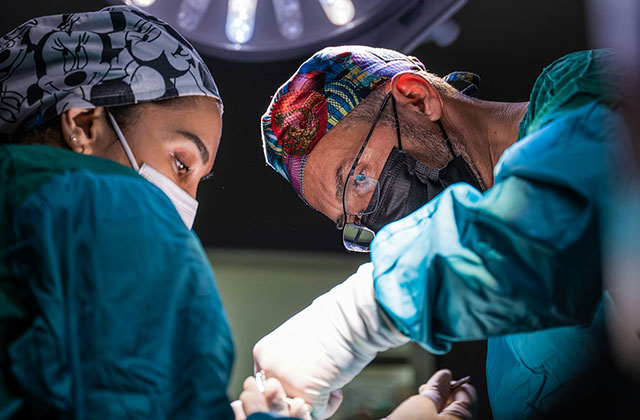
The operation to remove the chondromal body is performed using a minimally invasive arthroscopic method. In uncomplicated cases, this intervention does not take much time, is not associated with major trauma, and recovery is very fast.
How the operation is performed:
- Pain relief: spinal anesthesia or general anesthesia.
- Making several incisions up to 0.5 cm in the knee joint area.
- Installation of cannulas (expanders) that allow the removal of chondromal bodies.
- Insertion of an arthroscope and examination of the knee joint to identify all chondral bodies and assess the condition of intra-articular tissues (menisci, articular cartilage, synovial membrane).
- Extraction of chondromal bodies entirely or with preliminary fragmentation.
- A possible stage of the operation is partial synovectomy – partial removal of the synovial membrane if it is damaged and inflamed.
It happens that chondromous bodies elude the doctor, and he cannot grasp them to pull them out. In this case, an additional puncture with a needle may be required to fix the fragment, or the installation of an additional port for the introduction of a clamp with a serrated working surface.
Although very large chondromal bodies must be fragmented, doctors still try to avoid this procedure by removing them whole. This may require making a slightly larger incision than usual.
Some cartilaginous bodies are fused with the synovial membrane. In this case, they are first captured so that the joint mice do not escape after separation, and then cut off from the synovial membrane with an electric knife.
Sometimes it is necessary to restore the defect of the articular cartilage in the place where the fragment “fell off”. Mosaic autochondroplasty can be used for this purpose. The doctor takes fragments of cartilage from the non-load-bearing area of the knee joint to install them in the defect area. Although this procedure significantly increases the rehabilitation period, it prevents the development of osteoarthritis of the knee joints.
Rehabilitation after surgery
If the operation to remove chondromal bodies was performed as an isolated intervention, possibly also with synovectomy, then rehabilitation does not take much time. After a month, most people feel healthy, and after a month and a half, they can return to their usual active life.
Features of rehabilitation:
- On the day of surgery, elastic bandaging of the legs is indicated to prevent thromboembolic complications;
- massage and physiotherapy procedures begin on the same day;
- the next day you can begin therapeutic exercise: for now these are only isometric (without knee movements) contractions of the thigh muscles;
- From the third day after the operation, you can put weight on the operated leg;
- after 10-14 days, soft tissue wounds heal, stitches are removed, and you can walk without crutches;
- The period after two weeks is functional, and during this time a person performs exercises to restore volume, muscle strength, and knee mobility.


